By Jayden Malseed
They say a picture’s worth a thousand words, but we’re hoping these brightly coloured images can tell an even bigger story.
At first glance you may think the image below is part of an octopus tentacle, or maybe the underside of an alien spaceship from the 1996 movie Independence Day, or perhaps even something else entirely.
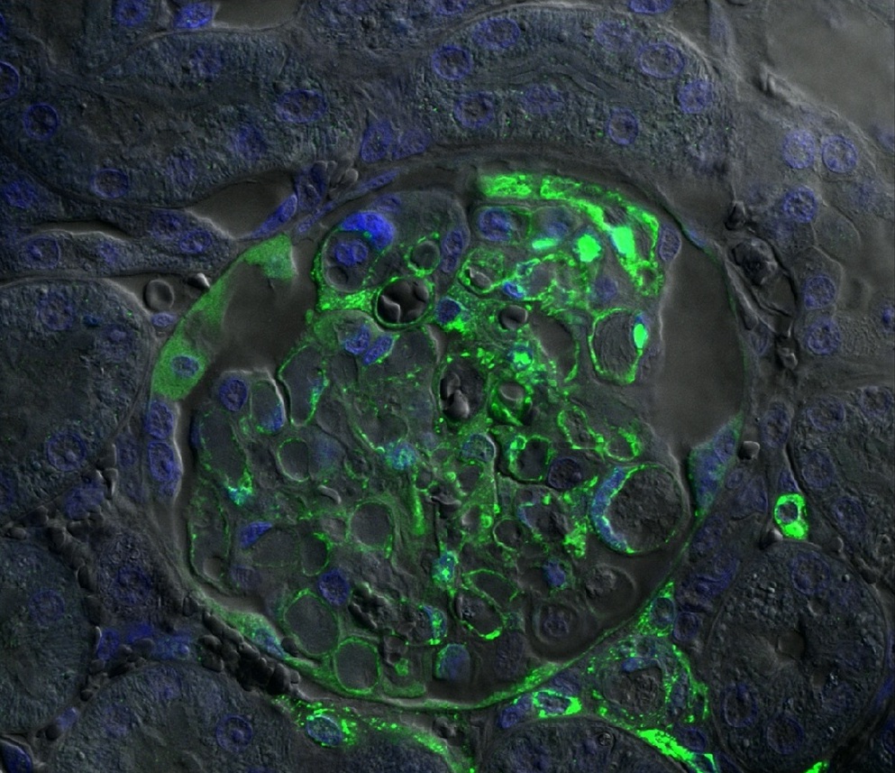
Is it an octopus? Is it a spaceship?…nah…it’s only 200 microns wide!
It’s actually a section from a ferret’s kidney that is infected with Hendra virus, and has been taken with a confocal microscope.
Now this isn’t just your ordinary microscope. Costing roughly $750 000, the microscope is designed to focus on fluorescent colours that have been ‘tagged’ to specific components, which then show up on a big computer screen, giving us these incredible pictures.
The green highlights are the cells that have been infected by Hendra, while the blue highlights are the cell nuclei. To create this picture an antibody is dyed fluorescent green, which then attaches to the viral proteins, effectively colouring it green.
The vital research, led by microscopist Dr Paul Monaghan, uses these images to study the cell biology of Hendra virus. The confocal microscope, which is located within the high containment facility at our Australian Animal Health Laboratory in Geelong Victoria, helps Paul and his team better understand the virus, and to be able to answer questions such as why it attacks certain cells, and what it does when it gets to a cell.
“We’re developing a deeper understanding of the virus by using the microscope and the images, and if we can pinpoint a specific stage in the virus lifecycle and say to ourselves ‘this is the point we need to stop it’ then that would be enormous”, Paul explained.
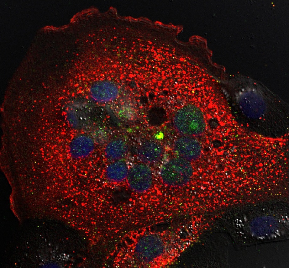
This is a confocal image of tissue culture taken 18 hours after inoculation with Hendra virus, and is about 100nm wide
The two images to the right are slightly different from the first. Where the first was a section from a kidney, these are taken from cells growing in tissue culture. We have also labeled two virus proteins: one red and one green.
They demonstrate how the Hendra virus has infected the cells, and after 14 hours has fused those cells together to form what is called a syncytium. The green/blue round circles are the nuclei – normally one in each cell – but the rest of the cell is relatively unaffected.
…and after 24 hours.
After 24 hours, the infection has progressed and newly made virus proteins are gathering at the edge of the cell (next to the black areas) to form new viruses. The red and green proteins are now together and can be seen as an irregular orange line at the edge of the cell.
These images allow Paul and his team to study the virus at different stages of its lifecycle, and and will be incredibly helpful for future research with Hendra virus and other related viruses that threaten the biosecurity of our animals, people and environment.
This research is part of our wider program of work on bats and the viruses they carry.