The Maia Mapper is our latest and most advanced instrument in our armoury in the search for minerals.
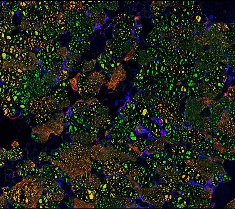
Characterisation image of natural mineral iron-oxide nodules. Taken with CSIRO-developed Maia x-ray imaging
Characterisation image of natural mineral iron-oxide nodules. Taken with CSIRO-developed Maia x-ray imaging
Did you know even ordinary rocks contain minuscule amounts of copper and gold? Ore deposits form when these precious elements become enriched by fluids flowing through the rocks. Mineral explorers rely on patterns in these elements to guide them to valuable ore deposits.
Thanks to a world-first innovation, we’ve made this process even easier. We have just built the world’s first high performance analysis tool that uses micro-X-ray fluorescence imaging to ‘illuminate’ trace elements of interest to mineral exploration and processing companies.
The new prototype, named the Maia Mapper, allows scientists to measure low concentrations of chemical elements with the precision of the thickness of a human hair in samples that are 50 cm long. This combination of sensitive, fine-scale and large area analysis – from the micro-to-metre scale – will transform our understanding of how elements are distributed around major ore deposits.
This new instrument builds upon our original innovation, the Maia detector, which was previously only available on high-end synchrotron facilities, like the Australian Synchrotron in Melbourne. Now researchers will be able harness the power of their very own Maia detector in their lab, with a slightly less powerful X-ray beam in place of the synchrotron.
How does the Maia Mapper work?
The Maia Mapper fires an X-ray beam at the rock sample but instead of seeing through it like a conventional medical X-ray, the beam gives the atoms enough energy so that they emit X-rays of their own. The Maia detector then measures the X-rays of each element present.
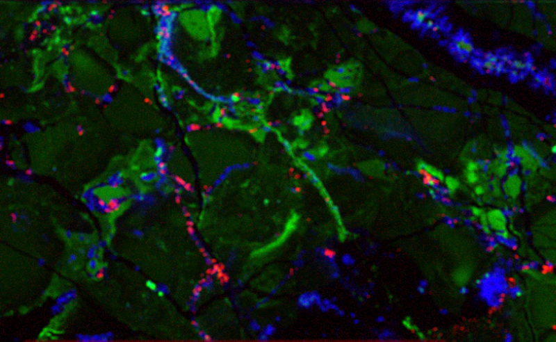
Imaging from the maia mapper showing different elements in blue
Cross section of a drill core from the Abra mineral deposit, Western Australia, showing three elements: copper (red), iron (green) and lead (blue).
‘Ore-some’ images
Our first ‘ore-some’ images from the Maia Mapper have been of rock core samples from the Abra deposit, Western Australia. We have been studying the rocks at Abra to help us understand ‘distal footprints’, which are markers that give us clues about deep mineral systems.
These beautiful images display a colourful palette of red, green and blue, which show the location and amount of copper, iron and lead respectively.
The highly detailed information will help solve a variety of mining industry problems like where the minerals are, what they mine first and how they get the valuable elements out of the rocks helping to streamline to whole process.
The Maia Mapper has been developed as part of the Advanced Resource Characterisation Facility project supported by the Science and Industry Endowment Fund (SIEF). The project aims to provide cutting edge analysis tools to benefit the minerals industry in a collaborative project between CSIRO, The University of Western Australia and Curtin University in Perth under the collaboration framework of the National Resources Science Precinct.

5th October 2016 at 6:53 pm
At what point do we refrain from relocating billions of tonnes of metals from the southern hemisphere before we change our axis?
5th October 2016 at 1:23 pm
While the statement “world’s first high performance analysis tool that uses micro-X-ray fluorescence imaging” may seem a little naive as there are numerous systems for XRF mapping available, the Maia Mapper brings together arguably the best technology for micro-focus X-ray source, achromatic focusing and real-time XRF detection, in the form of the R&D 100 Award winning, CSIRO-BNL developed, Maia detector system. The result is a high performance XRF mapping instrument particularly optimized for mineral samples, such as drill core segments to 500 mm imaged with micron scale detail.