
Mmm . . . lovely! A hot Indian curry is simmering away on the stove on a wintry night. The smell of spices fills the kitchen. One of the spices is turmeric, from the ginger family. Its vibrant yellow colour comes from the compound curcumin which is finding a use in clinical tests for Alzheimers disease (AD).
Who knew? Soon everyone will! We’re presenting our research this week at a major conference in Copenhagen, AAIC2014.
A clinical trial of the spice-infused eye test is being led by our own Dr Shaun Frost and team, with WA’s Edith Cowan University, US company NeuroVision Imaging, and the McCusker Alzheimer’s Research Foundation in Perth. Several hundred volunteers have taken part. They include healthy people, mildly cognitively impaired people and patients with AD. It’s all part of the Australian Imaging Biomarkers and Lifestyle study of Aging (AIBL)
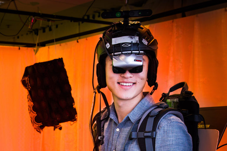
The trial asks volunteers to come along to two visits for retinal fluorescence imaging, ie an eye scan. This is quick and painless. Patients sit in front of a specialised camera and a photo is taken of the retina at the back of their eye.

Between visits, volunteers eat some curcumin which binds to beta-amyloid plaques, the sticky proteins that indicate Alzheimers, and fluoresces. The plaques (if there are any) show up in the eye scans as bright spots which can be counted and measured. The data is then used to calculate a special number for each patient, a retinal amyloid index (RAI), and compared between healthy, mildly cognitively impaired and AD patients.
Encouragingly, as we announced this week, early results show the amount of plaque in the retina closely mirrors the amount in the brain. If confirmed, retinal imaging may be the beginnings of an easy, non-invasive test for early detection of AD. Combined with results of cognitive tests and other markers it could help doctors diagnose AD more confidently.
Eye scans like this also find plaques when they’re smaller than the ones in brain scans, potentially finding signs of AD earlier – maybe up to 20 years before cognitive symptoms appear. If diagnosed, AD patients could start treatment sooner and have regular eye scans to see which treatments work best for them.
Brain imaging on the cloud
From curry to the cloud. More research presented this week is about more accurately interpreting brain images sometimes used to diagnose AD.
To get a brain scan, a patient lies on a bed in a large machine like a Magnetic Resonance Imaging (MRI) or Positron Emission tomography (PET) scanner. These machines record a series of images through the brain, which are then visually checked by a radiologist who compiles a report for the patient’s doctor.
This visual inspection can be subjective, tedious and time consuming. But recent advances in scientific computing and machine learning allows systems to accurately measure features of the 3D scan, such as brain size or concentration of a tracer molecule, that support a diagnosis.
Using these techniques, a new trend is emerging for improving radiologists’ productivity. Scanners and specialised medical software can report quantitative values and compare them to the values expected for normal, healthy patients – just like blood test results from a pathology lab do.
Our researchers, led by health imaging specialist Associate Prof Olivier Salvado, have just released a new cloud computing application, MILXCloud, that automatically delivers standardised radiology reports.

Users will be able to upload a PET scan and within 15 minutes be emailed a one page quantitative report showing a diagram of the brain with colour coded values compared with what’s normal. This data will help support diagnosis by the radiologist and enhance delivery of eHealth services.
Whether it’s curry or the Cloud, the future of Alzheimer’s detection sure looks bright.
6th August 2014 at 8:54 am
If you start conducting any trials on the retinal scanning for AD detection in Victoria, pick me! With a mother and grandfather both having AD, I worry that I am going the same way everytime my memory fails me – which is increasingly often!